
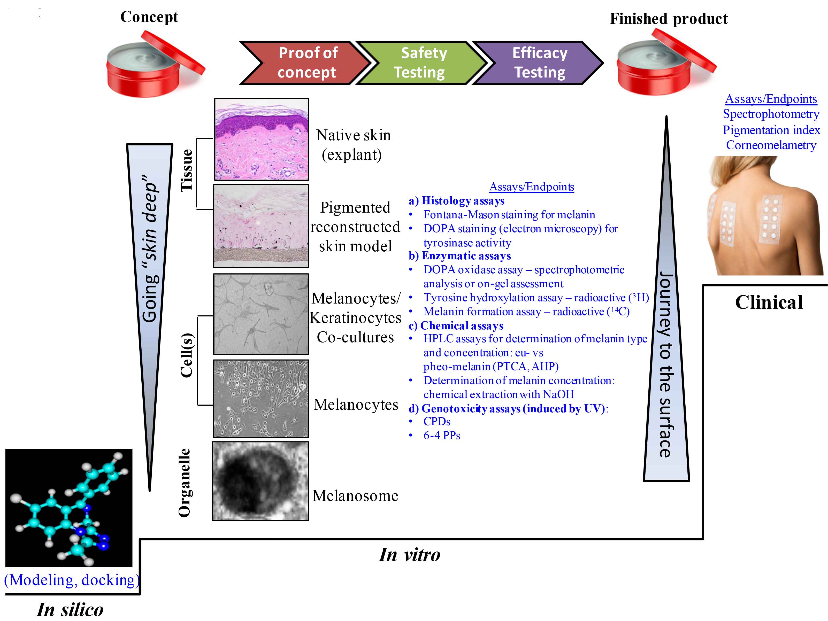
Figure 5: Going “skin deep” and the journey back to the skin surface: available assays and testing strategies integrated into the skin care products manufacturing framework. AHP, aminohydroxyphenylalanine; CPD, cyclobutane pyrimidine dimers; DOPA, L-3,4-dihydroxyphenylalanine; HPLC, High performance liquid chromatography; NaOH, sodium hydroxide; PPS, photoproducts; PTCA, pyrrole-2,3,5-tricarboxylic acid; UV, ultraviolet light. Panel “Pigment reconstructed skin model” represents the MelanoDerm™ tissue model MEL-300-A after two weeks in culture; Fontana-Mason staining, magnification 10x; courtesy to MatTek Corporation, Ashland, MA, USA; Panel “Melanocytes/Keratinocytes Co-cultures” reproduced from Kumar R. et al., Ind. J. Dermatol. Venereol. Leprol., 78, 599-604 (2012);1 Copyright Wolters Kluwer Medknow Publications; Panels “Melanocytes” and “Melanosome” reproduced (with slight modifications) from Costin et al., J. Cell. Sci., 116, 3203-3212 (2003); Copyright The Company of Biologists Ltd.2
References
1.
R. Kumar, D. Parsad, A. Kanwar, and D. Kaul, Development of melanocyte-keratincoyte co-culture model for controls and vitiligo to assess regulators of pigmentation and melanocytes, Ind. J. Dermatol. Venereol. Leprol., 78, 599-604 (2012).
2.
G.E. Costin, J.C. Valencia, W.D. Vieira, M.L. Lamoreux, and V.J. Hearing, Tyrosinase processing and intracellular trafficking is disrupted in mouse primary melanocytes carrying the underwhite (uw) mutation. A model for oculocutaneous albinism (OCA) type 4, J. Cell. Sci., 116, 3203-3212 (2003).