
by The Cosmetic Chemist Staff
Poor oral care health can result in a number of unfavorable conditions in the oral cavity that are principally caused by bacteria. There has been considerable interest in the cosmetics and toiletries industry to design and invent products in the form of dentifrices for the treatment and prevention of these ailments. Plaque and calculus (tartar) are principle concerns as well, as they can lead to more serious conditions such as the formation of caries, commonly known as cavities, or the development of gingivitis (inflammation of the gum tissue). Dental plaque is a yellow biofilm film formed on the surface of the tooth enamel due to bacterial colonization (see photograph below). There can be as many 1,000 bacterial species present and plaque owes its bacterial diversity due to the lack of a biological self-cleansing system.1 Initially, the biofilm is soft in nature; however, after several days it begins to harden forming tartar, also known as calculus.
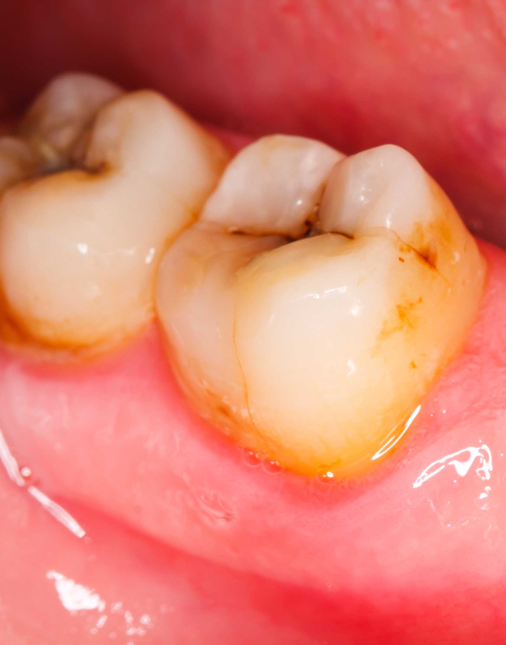
Photograph of the oral cavity illustrating the formation of a yellow biofilm, known as dental plaque, on the surface of the tooth enamel.
Dental biofilms represent a significant threat to oral health as many of the bacteria present in dental plaque are cariogenic—they cause demineralization of the tooth. Typically, such bacteria are acidogenic (they produce organic acids) and aciduric (they are able to survive in acidic environments). The most prevalent of these bacteria are Lactobacillus species, Streptococcus sobrinus, and Streptococcus Mutans.2 Their degradative activity occurs as a result of fermentation of dietary carbohydrates (e.g., sucrose) into formic, acetic, and propionic acids, which makes the tooth more susceptible to the perils discussed above.
The enamel pellicle contains a protective layer of proteins (statherin, proline-rich proteins, and mucinous proteins from saliva) that bind to hydroxyapatite. In addition to these proteins, there are other salivary proteins (e.g., cystatins, histatins, lysozyme, amylase, lactoferrin, lactoperoxidase, carbonic anhydrate, etc.) that also deposit on the surface of the enamel pellicle and protect it from metabolic byproducts of bacteria that comprise the biofilm.3 These proteins have a variety of functions in protecting the tooth, such as inhibiting the adhesion of microorganisms and even helping the tooth remineralize by binding important trace elements, such as calcium.
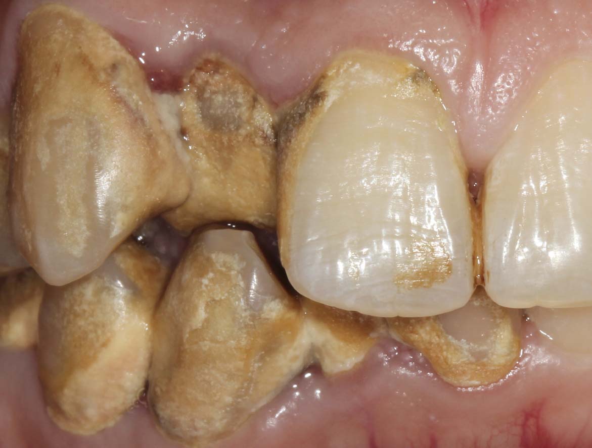
Formation of supragingival calculus on the tooth surface. In the image, heavier deposits of calculus are observed on the subject’s right teeth (left side of the photograph).
Dental calculus, commonly known as tartar, results from the crystallization of dental plaque. As already noted, plaque exists in the form of a soft biofilm, which hardens due to the accumulation of minerals from saliva, such as calcium phosphate. Often, calculus is further classified as supragingival or subgingival, which indicates the location where it is found—supragingival calculus occurs on the crown of the tooth while subgingival forms lower in pockets of the gingiva. Supragingival calculus has a mineral content of about 37%, and also contains proteins and lipids, which are also believed to be an integral part of calculus formation.4 For illustration, the photograph above contains teeth subjected to calculus formation. It should be noted that calculus cannot be removed simply by brushing and other normal oral hygiene routines. Usually it requires special dental tools such as a traditional or ultrasonic tooth scaler since it is so tightly bound to the tooth surface. Calculus buildup, however, can be prevented when suitable dentifrices are utilized on a routine basis.
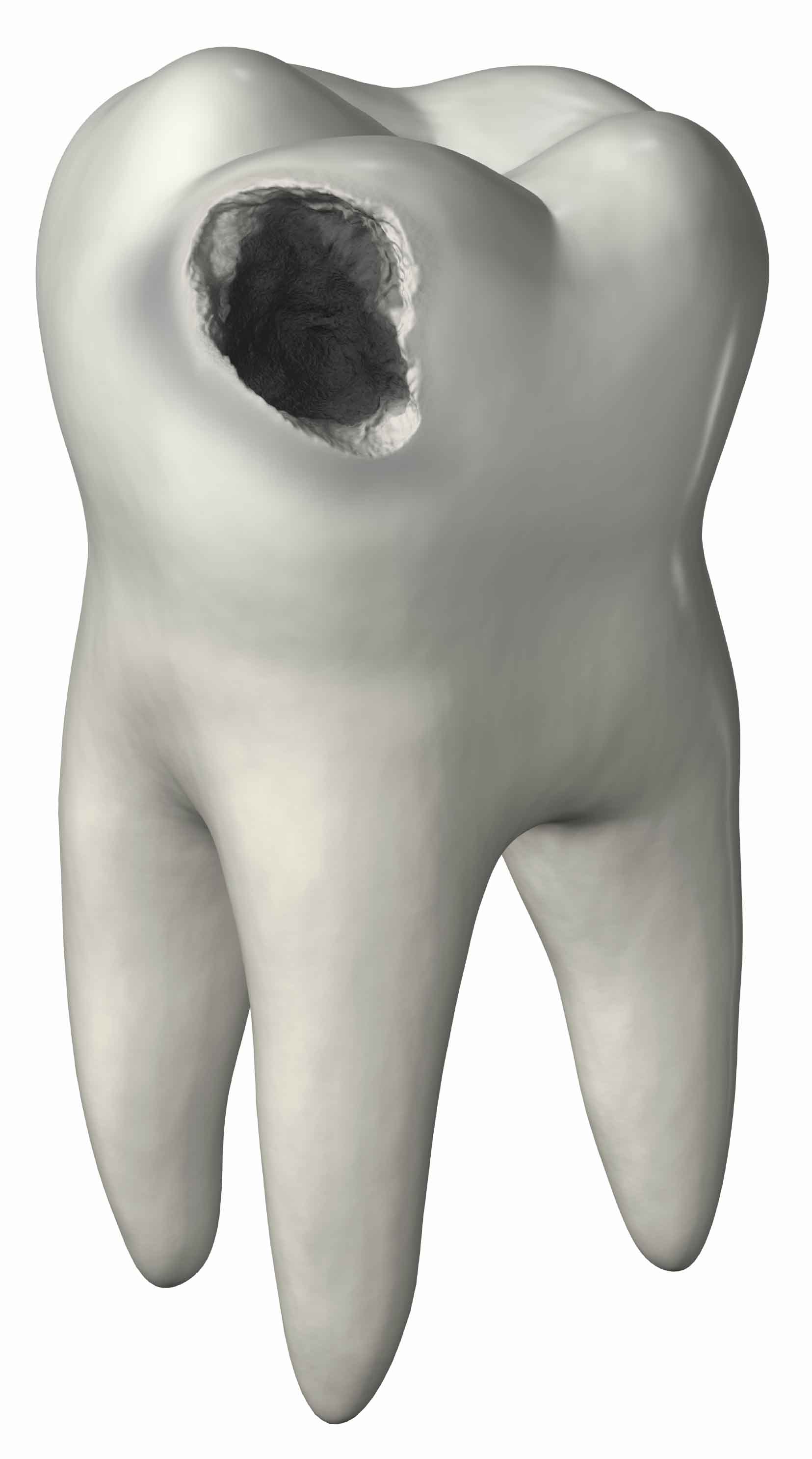
Illustration of a carious human tooth (molar). Demineralization can result in a visible cavity in the tooth structure.
Dental caries, commonly known as cavities, form as a result of demineralization of the tooth structure. The acidic environment that prevails during the presence of plaque and calculus ultimately leads to damage of the tooth’s enamel coating and can extend into the interior dentin structure. Carie formation is usually detected in the early stages during routine dental checkups. However, there are instances when visible pits or holes can be found in the tooth, such as that shown in the illustration above. Gingivitis is a type of periodontal disease resulting in inflammation and infection of the gum tissue (gingiva). It is a degenerative disease that is caused by the long-term effects of dental plaque and calculus deposits on the teeth. The bacteria associated with plaque and calculus, along with their metabolites, infect the gum tissue eventually leading to the degeneration of tissue that supports the tooth, such as periodontal ligaments and alveolar bone tissue of the tooth socket.
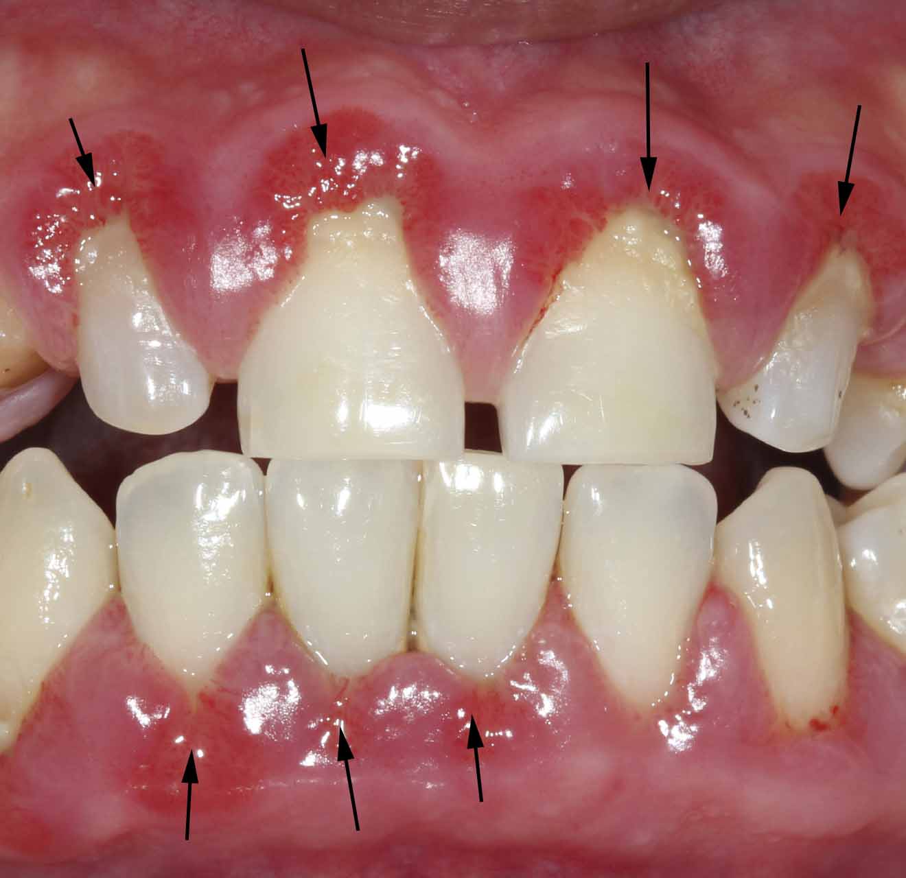
Front-facing photograph of the oral cavity illustrating the development of gingivitis (marked by arrows) in regions close to the base of the teeth.
Gingivitis can progress into a more serious, but related condition, known as periodontitis or gum disease. It is the irreversible breakdown of gingiva and bone tissue surrounding the tooth. Eventually, it leads to tooth loss. Even graver, it can increase the risk of heart disease and stroke and suppress the immune system. Periodontitis is mostly caused by poor oral hygiene and can be avoided by proper brushing and flossing.
References
1.
J. ten Cate, Biofilms, a new approach to the microbiology of dental plaque. Odontology, 94(1), 1-9 (2006)
2. F. García-Godoy, and M. Hicks, Maintaining the integrity of the enamel surface: The role of dental biofilm, saliva and preventive agents in enamel demineralization and remineralization. J. Am. Dent. Assoc., 139(suppl), 25S-34S (2008).
3. A. Van Nieuw Amerongen, J. Bolscher, and E. Veerman, Salivary proteins: protective and diagnostic value in cariology? Caries Res., 38, 247-253 (2004).
4. Y. Jin, and H.-K. Yip, Supragingival calculus: formation and control. Crit. Rev. Oral Biol. Med., 13(5), 426-441 (2002).