
by The Cosmetic Chemist Staff
The human tooth is a complex morphological structure, in which part of the tooth is exposed, while a considerable amount is embedded in the gums (gingiva) and extends into the jaw bone (mandibula). An illustration of tooth and its component parts is provided below, and can be subdivided into three components: root, neck, and crown.
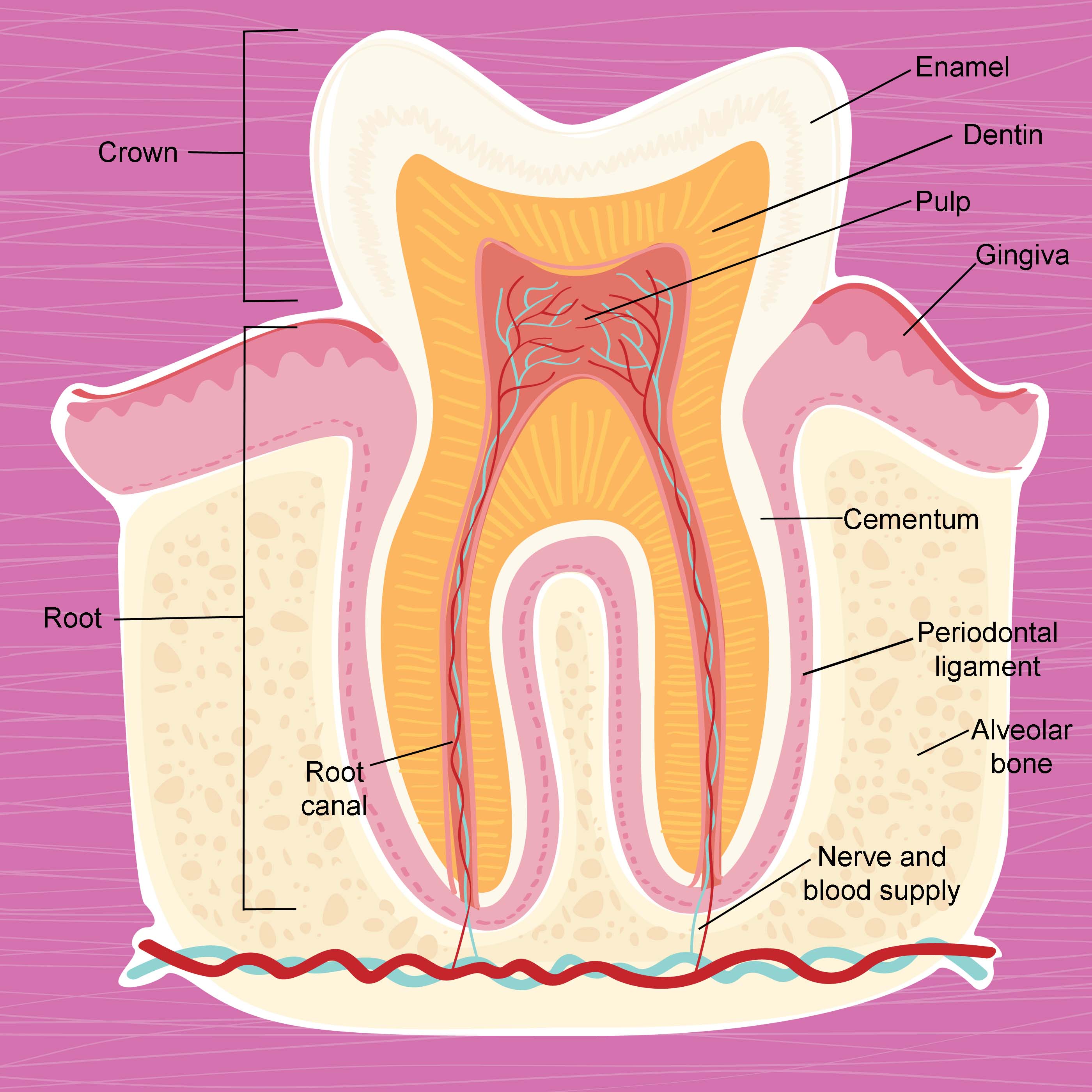
Tooth anatomy.
As already mentioned, the root is embedded in the bone mass of the mandibula—the alveolar bone. The relatively long root structure also contains nerve tissue that extends up until the pulp cavity, which also contains pulp and blood vessels. Odontoblast cells in the pulp are responsible for forming dentin. Nutrients are delivered to this region via the blood supply and allow the tooth to sustain itself as a living tissue. The presence of nerves allows for the sensation of pain, which is often caused by extreme temperatures, and is especially noticeable when drinking cold or hot beverages. The root canal (also known as the pulp canal) is the narrow conduit starting at the base of the root and extending up until the pulp cavity, which provides access for vascularization and innervation. Problems in the pulp cavity, such as infection, often require root canal therapy.
Surrounding the root structure, the cementum provides a biological adhesive layer between the main body of the root and the periodontal ligament. It is composed of fibrous tissue, which firmly attaches the teeth to the jaws. One end of the fiber bundle is connected to the cementum and the other into the alveolar socket, which is the connection point to the jaw. The neck of the tooth describes the area where the gingiva (gum tissue) is located and the tooth becomes exposed to the buccal cavity (the region of the oral cavity that is bound by the lips, cheeks, and gums).
Finally, the crown is the visible portion of the tooth and contains a hard, crystalline outer layer of enamel that protects the interior structure of the tooth by preventing demineralization. It also facilitates mastication, as it is a very smooth surface. Since enamel is a hard structure, it is also very brittle. Enamel is a translucent layer that is susceptible to damage due to eating sugary foods or drinking acidic beverages and is not like other bone tissue, which the body can self-heal. Staining of teeth usually occurs on the enamel layer and is caused by consuming beverages, such as tea and coffee, as well as by smoking tobacco or even poor oral hygiene. Normal tooth color is not pure white, but a slight off-white, gray, or even yellow color. Below the enamel, most of the tooth structure is composed of dentin, a bone-like substance that provides teeth with their characteristic color.
At the chemical level, tooth is principally comprised of crystalline calcium phosphate, which exists in the mineral form as hydroxyapatite and has the molecular formula Ca5(PO4)3(OH). Enamel, which again is very hard and brittle, contains relatively high levels of hydroxyapatite (ca. 92-94%) as well as much lower quantities of water (2-3%), carbonate (2%), trace elements (1%) (e.g., sodium, magnesium, potassium, chloride, zinc), fluoride (0.01-0.05%), and proteins and lipids (<1%).1 Dentin, on the other hand, contains much less hydroxyapatite and is also comprised of collagen and other organic matter (35-50%).
Reference
1.
J. Hicks, F. Garcia-Godoy, and C. Flaitz, Biological factors in dental caries enamel structure and the caries process in the dynamic process of demineralization and remineralization (part 2). J. Clin. Pediatr. Dent., 28(2), 119-124 (2004).