
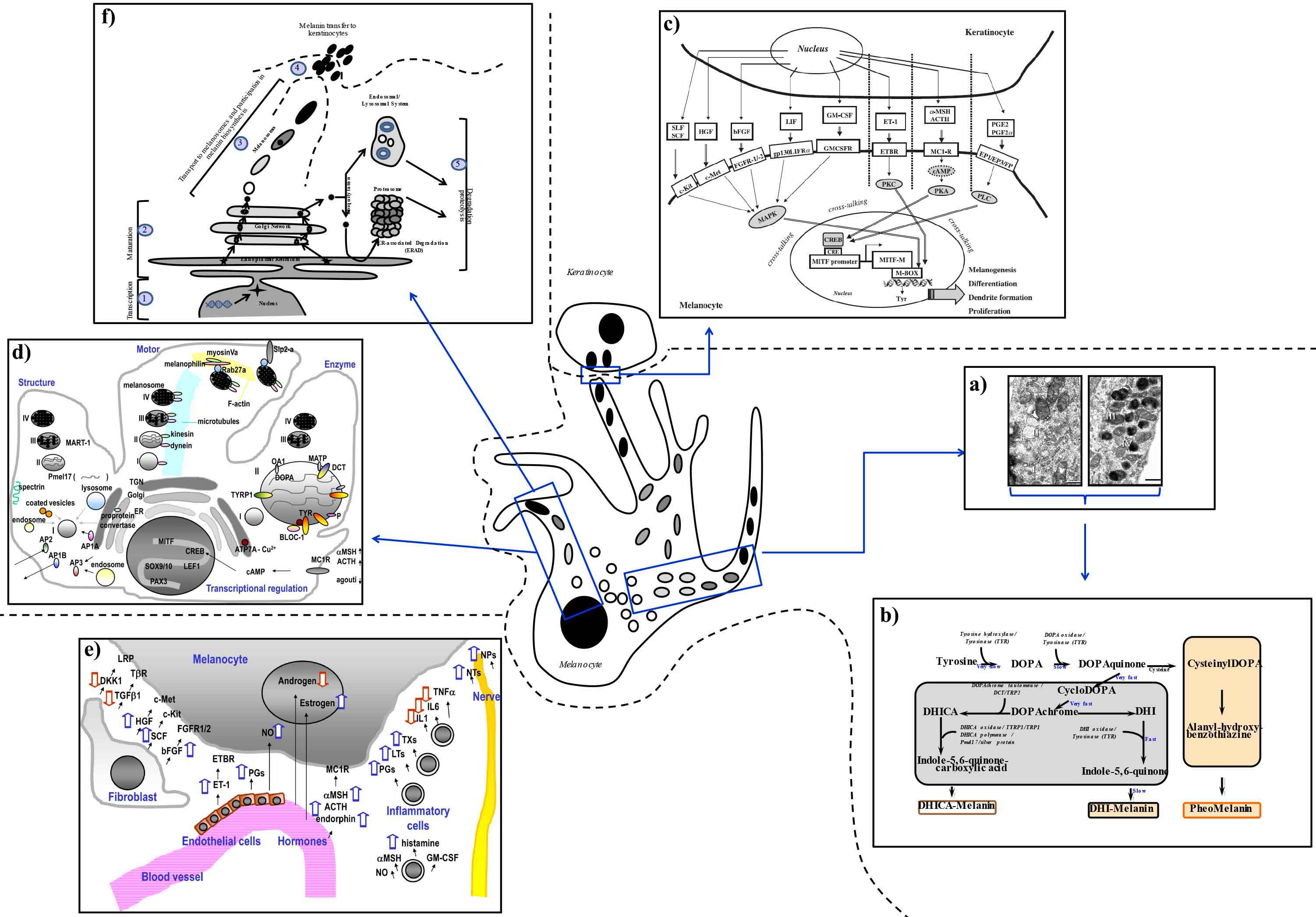
Figure 4: Positioning the melanocyte in its physiological environment.
(a) Stages of melanosomes development. Images reproduced (with slight modifications) with permission from Costin et al., J. Cell. Sci., 116, 3203-3212 (2003); Copyright The Company of Biologists, Ltd.1
(b) Melanin biosynthesis pathway. Abbreviations: DCT, Dopachrome Tautomerase; DHI, 5,6-dihydroxyindole; DHICA, DHI-2-carboxylic acid; DOPA, L-3,4-dihydroxyphenylalanine; TRP2, Tyrosinase related protein 2 (also known as DCT); TYR, Tyrosinase; TYRP1, Tyrosinase related protein 1 (also known as TRP1).
(c) Extrinsic factors regulating melanocyte functions: UV action on melanocytes and keratinocytes. After exposure of the skin to UV light, p53 initiates the release by keratinocyte of melanogenic paracrine growth factors and cytokines: adrenocorticotropic hormone (ACTH),2 α-melanocyte stimulating hormone (α-MSH),2,3 endothelin-1 (ET-1),4-6 stem cell factor (SCF),7 hepatocyte growth factor (HGF),8 basic fibroblast growth factor (bFGF),5,6 leukemia inhibitory factor (LIF),9 and granulocyte macrophage colony-stimulating factor (GM-CSF).10,11 Prostaglandin PGE2 and PGF2a are synthesized and released by human keratinocytes following the exposure to UV and subsequently stimulate the dendritogenesis of melanocytes.12 These factors interact with their corresponding receptors on melanocytes, and induce melanocyte activation, with subsequent stimulation of microphtalmia-associated transcription factor (MITF)13,14 and its downstream targets, the melanogenic enzymes Tyrosinase (TYR), Tyrosinase related protein 1 (TYRP1), and Dopachrome tautomerase (DCT) ending with the biosynthesis of melanin. MITF, the master regulator of melanogenic pathway, is activated through three signaling pathways regulated by cyclic Adenosine monophosphate (cAMP),15,16 Protein kinase C (PKC),14,17,18 phospholipase C (PLC)12 and mitogen-activated protein kinase (MAPK)14,18 to subsequently induce proliferation and differentiation of melanocytes. Reproduced with permission from G.E. Costin and V.J. Hearing, FASEB J., 21, 1-19 (2007); Copyright Federation of American Societies for Experimental Biology (FASEB).13
(d) Intrinsic factors regulating the melanocyte: enzymatic, structural, transcriptional, and motility-related factors. Reproduced from Y. Yamaguchi Y. and V.J. Hearing, Biofactors, 35, 193-199 (2009); Copyright John Wiley and Sons.19 Abbreviations: ACTH, adrenocorticotropic hormone; ATP7A (copper-transporting P-type ATPase or Menkes protein); AP, adaptor protein; BLOC, biogenesis of lysosome-related organelles complex 1; cAMP, cyclic Adenosine monophosphate; CREB, cAMP response element binding protein; DCT, Dopachrome tautomerase; ER, endoplasmic reticulum; MART, melanoma antigen recognized by T-cells 1; MATP, membrane-associated transporter protein; MC1R, melanocortin 1 receptor; MITF, Microphthalmia-Associated Transcription Factor; MSH, melanocyte stimulating hormone; OA, ocular albinism; PAX, paired box; PG, prostaglandin; SOX, SRY-Box; TCF/LEF, T-cell factor/lymphoid enhancer factor; TGN, Trans Golgi Network.
(e) Factors regulating the communication between melanocytes and other cell populations residing in the skin. Reproduced from Y. Yamaguchi and V.J. Hearing, Biofactors, 35, 193-199 (2009); Copyright John Wiley and Sons.19 Abbreviations: ACTH, adrenocorticotropic hormone; bFGF, basic fibroblast growth factor; c-Kit, mast/stem cell growth factor receptor (SCFR) or tyrosine-protein kinase Kit or CD117; c-Met, Hepatocyte growth factor receptor; DKK1, Dickkopf; ET, endothelin; ETBR, endothelin B receptor; FGFR, fibroblast growth factor receptor; GM-CSF, granulocyte macrophage-colony stimulating factor; HGF, hepatocyte growth factor; IL, interleukin; LRP, lipoprotein receptor-related protein; LT, leukotriene; MC1R, melanocortin 1 receptor; MSH, melanocyte stimulating hormone; NO, nitric oxide; NP, neuropeptide; NT, neurotrophin; PG, prostaglandin; TGF, transforming growth factor.
(f) Key steps in TYR processing, maturation, acquisition of enzymatic activity, and degradation and melanin transfer to keratinocytes that can be affected by melanogenesis modifiers (see also Table 1 and Table 2), and available assays and endpoints.
References
1. G.E. Costin,
J.C. Valencia, W.D. Vieira, M.L. Lamoreux, and V.J. Hearing, Tyrosinase processing and intracellular trafficking is disrupted in mouse primary melanocytes carrying the underwhite (uw) mutation. A model for oculocutaneous albinism (OCA) type 4, J. Cell Sci., 116, 3203-3212 (2003).
2. A.T. Slominski, B. Roloff, B. Zbytek, E.T. Wei, K. Fechner, J. Curry, and J. Wortsman, Corticotropin releasing hormone and related peptides can act as bioregulatory factors in human keratinocytes, In Vitro Cell Dev Biol Anim., 36, 211-216 (2000).
3. H. Holzmann, P. Altmeyer, L. Stöhr, and G.N. Chilf, Modification of alpha-MSH by UVA irradiation of the skin, Der Hautarzt, 34, 294-297 (1983).
4. G. Imokawa, T. Kobayashi, M. Miyagishi, K. Higashi, and Y. Yada, The role of endothelin-1 in epidermal hyperpigmentation and signaling mechanisms of mitogenesis and melanogenesis, Pigment Cell Res., 10, 218-228 (1997).
5. C.S. Wu, C.L. Yu, C.S. Wu, C.C. Lan, and H.S. Yu, Narrow-band ultraviolet-B stimulates proliferation and migration of cultured melanocytes, Exp. Dermatol., 13, 755-763 (2004).
6. M. Brenner, K. Degitz, R. Besch, and C. Berking, Differential expression of melanoma-associated growth factors in keratinocytes and fibroblasts by ultraviolet A and ultraviolet B radiation, Br. J. Dermatol., 153, 733-739 (2005).
7. A. Hachiya, A. Kobayashi, Y. Yoshida, T. Kitahara, Y. Takema, and G. Imokawa, Biphasic expression of two paracrine melanogenic cytokines, stem cell factor and endothelin-1, in ultraviolet B-induced human melanogenesis, Am. J. Pathol., 165, 2099-2109 (2004).
8. M. Mildner, V. Mlitz, F. Gruber, J. Wojta, and E. Tschachler, Hepatocyte growth factor establishes autocrine and paracrine feedback loops for the protection of skin cells after UV irradiation, J. Invest. Dermatol., 127, 2637-2644 (2007).
9. R.C. McKenzie, Ultraviolet radiation B (UVB)-induction of Leukaemia Inhibitory Factor (LIF) in human keratinocytes, Photodermatol. Photoimmunol. Photomed., 17, 284-285 (2001).
10. G. Imokawa, Y. Yada, M. Kimura, and N. Morisaki, Granulocyte/macrophage colony-stimulating factor is an intrinsic keratinocyte-derived growth factor for human melanocytes in UVA-induced melanosis, Biochem J., 313, 625-631 (1996).
11. D.S. Kim, H.J. Kim, K.H. Choi, J.H. Chung, K.H. Kim, and K.C. Park, UVB-induced GM-CSF production is suppressed by dexamethasone in HaCaT cells, Photodermatol. Photoimmunol. Photomed., 17, 121-125 (2001).
12. G. Scott, S. Leopardi, S. Printup, N. Malhi, M. Seiberg, and R. Lapoint, Proteinase-activated receptor-2 stimulates prostaglandin production in keratinocytes: analysis of prostaglandin receptors on human melanocytes and effects on PGE2 and PGF2alpha on melanocyte dendricity, J. Invest. Dermatol., 122, 1214-1224 (2004).
13. G.E. Costin and V.J. Hearing, Human skin pigmentation: melanocytes modulate skin color in response to stress, FASEB J., 21, 976-994 (2007).
14. T. Hirobe, How are proliferation and differentiation of melanocytes regulated? Pigment Cell Melanoma Res., 24, 462-478 (2011).
15. Y. Mizutani, N. Hayashi, M. Kawashima, and G. Imokawa, A single UVB exposure increases the expression of functional KIT in human melanocytes by up-regulating MITF expression through the phosphorylation of p38/CREB, Arch. Dermatol. Res., 302, 283-294 (2010).
16. T. Passeron, J.C. Valencia, C. Bertolotto, T. Hoashi, E. Le Pape, K. Takahashi, R. Ballotti, and V.J. Hearing., SOX9 is a key player in ultraviolet B-induced melanocyte differentiation and pigmentation, Proc. Natl. Acad. Sci. U.S.A., 104, 13984-13989 (2007).
17. H.Y. Park, C. Wu, L. Yonemoto, M. Murphy-Smith, H. Wu, C.M. Stachur, and B.A. Gilchrest, MITF mediates cAMP-induced protein kinase C-beta expression in human melanocytes, Biochem. J., 395, 571-578 (2006).
18.
K. Liu, D. Yu, Y.Y. Cho, A.M. Bode, W. Ma, K. Yao, S. Li, J. Li, G.T. Bowden, Z. Dong, and Z. Dong, Sunlight UV-induced skin cancer relies upon activation of the p38 signaling pathway, Cancer Res., 73, 2181-2188 (2013).
19. Y. Yamaguchi and V.J. Hearing, Physiological factors that regulate skin pigmentation, Biofactors, 35, 193-199 (2009).