
By Jonathan Hadgraft,† Elizabeth Mauldin,†† and Gopinathan K. Menon†††
March 15, 2017
†Emeritus Professor of Biophysical Chemistry at The School of Pharmacy, University of London, London, UK
††Associate Professor of Pathology/Dermatology, University of Pennsylvania School of Veterinary Medicine, Philadelphia, PA, USA
†††California Academy of Sciences, San Francisco, CA, USA
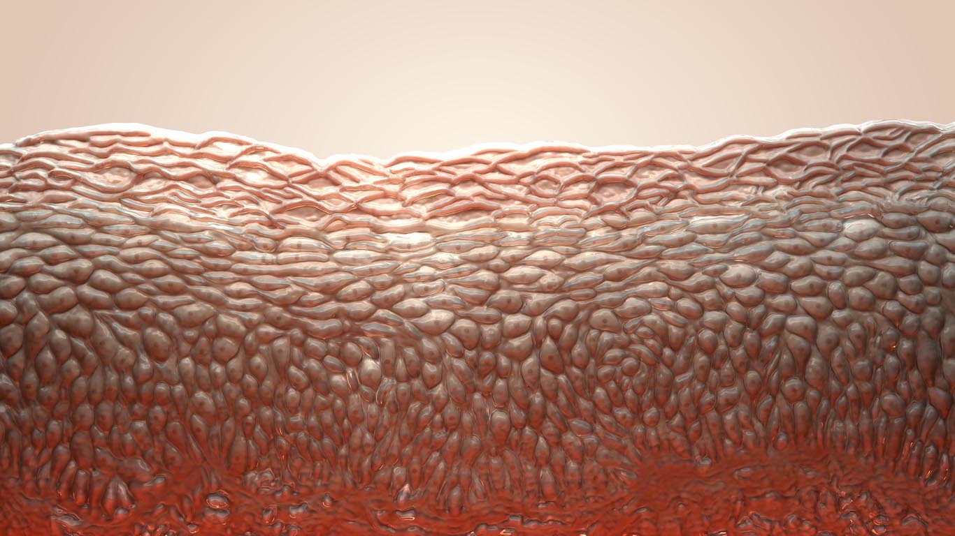
The role of an "acid mantle" of skin in maintaining the permeability barrier by creating a milieu that aids in the processing of secreted epidermal lamellar body contents seems to be widely accepted by skin biologists.1-4 The origin and maintenance of an acid pH of the stratum corneum and its significance for orderly exfoliation of corneocytes, as well as providing an antimicrobial barrier, have also been been addressed in the literature.5-7 Largely, the acid pH is measured using specialized instruments (e.g., a skin pH meter with a flat electrode) or evaluated by sophisticated microscopic techniques such as two-photon fluorescence lifetime imaging.8 We would like to revive discussion of the concept of pH measurement as well as examine if the "acid mantle" is truly a universal feature underlying the skin barrier.
Let us start with the basic tenets. The pH of the skin surface is generally regarded as being around pH 4. But, what does this mean? Does a sheet of paper or the computer screen on which you are reading this article have a pH? The second tenet is that viable tissue is approximately pH 7.
As we know from our training in chemistry, pH is defined as the negative logarithm of the hydrogen ion activity (often approximated as the concentration) in aqueous solution. The skin surface is certainly not aqueous. The general view of the pH of the skin is that if a drop of water is placed on the skin and the pH of that drop is measured then it will be around pH 4. This phenomenon is thought to emanate from the free fatty acids that comprise the skin lipids (including sebum),lactic acid, and urocanic acid.
However, the pH that is measured using a flat electrode will depend on the volume of the drop of water and the area over which that drop of water is placed. How is this standardized, and, in any case does it mean anything? A similar line of reasoning could be applied to a piece of paper or computer screen. However, it would be farcical to think that such deduction is meaningful. It is generally thought that a pH gradient exists across the skin. How do we define the pH in the stratum corneum? How much free water is present in the stratum corneum? What is the meaning of pH in bound water and how would we measure it?
In the personal care industry, where topical products are designed to have compatibility with the skin (especially terms of pH), a controversial issue is the meaning of pH when dealing with a gel, cream, or ointment. Remember that pH is usually measured using a glass electrode and it is determined in an aqueous solution. If the solution is not aqueous, e.g. an ethanol-water mixture, the glass electrode will only provide an estimate of the apparent pH. In addition, it is not a simple matter to take a glass electrode, which is stored in aqueous buffer, and place it in the ethanol-water mixture. The glass electrode needs to be conditioned by equilibrating it in the solution whose apparent pH is to be measured.
Technically, there cannot be a pH of a cream or ointment. It is possible to talk about the pH of the aqueous phase of a semi-solid preparation, but this means that the pH needs to be measured before the two-phase system is manufactured, or by separating the phases after manufacture.
While the basic issue of pH measurement is debated, in panelist studies in personal care and clinical investigations, skin surface pH is measured using a pH meter equipped with a flat electrode that is specifically designed for this purpose, such as that supplied by Courage + Khazaka.9 Nagashima and coworkers recently described a dry electrode based pH measurement, and compared their values with those obtained with the Courage + Khazaka instrument.10 They surmise that the conventional pH electrode requires water, and that their dry probe measures different pH values, and conclude that this non-invasive method could affect other factors that determine skin pH. Notwithstanding these differences, measurements with microscopic techniques, such as two-photon fluorescence lifetime imaging using pH sensitive fluorescent dyes, indicate that acidic pH exists in the stratum corneum extracellular domains of murine skin.8 Experimental studies on murine models using "super acids" (that enhance barrier recovery following its disruption) and super base compounds (which retard the barrier recovery) have emphasized a key role for the acid milieu in stratum corneum permeability and barrier homeostasis. Several clinical studies have also reported that the skin surface pH is altered as a result of aging11and diseases,12 although the evidence appears not to be as strong or convincing as in the animal studies. In a study involving non-insulin dependent diabetics, skin pH was significantly higher in the intertriginous zones of diabetics compared to intertriginous zones of healthy individuals.13 Interestingly, there was no difference in forearm pH of the two groups.14
Let us disregard for a while the issue of how pH is measured and focus on the pH variations when measurements are made with the flat electrode instruments used for skin measurements. In the course of discussions with a veterinary researcher (Didier Pin), one of us (GM) learned that several domesticated animals have a skin surface pH above 6, and in fact may be as high as pH 8 or 9. Although publications in veterinary journals have documented this deviation from an acid mantle, researchers in the skin barrier field continue to take for granted that an acid pH is a universal feature.15,16 Yet, dogs, cattle, and goats (which we believe possess a functional permeability barrier) do not have an acid mantle; as described by studies using bioinstrumentation measurements. We would like to invite the views of readers on this topic, with the hope of resolving these confounding issues.
References
1. J.W. Fluhr and P.M. Elias, Stratum corneum pH: Formation and Function of the 'Acid Mantle', Exog. Dermatol., 1, 163-175 (2002).
2. J.W. Fluhr, P.M. Elias, M.Q. Man, M. Hupe, C. Selden, J.P. Sundberg, E. Tschachler, L. Eckhart, T.M. Mauro, and K.R. Feingold, Is the filaggrin-histidine-urocanic acid pathway essential for stratum corneum acidification? J. Invest. Dermatol., 130, 2141-2144 (2010).
3. J.P. Hachem, D. Crumrine, J. Fluhr, B.E. Brown, K.R. Feingold, and P.M. Elias, pH directly regulates epidermal permeability barrier homeostasis, and stratum corneum integrity/cohesion, J. Invest. Dermatol., 121, 345-353 (2003).
4. M.H. Schmid-Wendtner and H.C. Korting,
The pH of the skin surface and its impact on the barrier function, Skin Pharmacol. Physiol., 19, 296-302 (2006).
5. H. Lambers, S. Piessens, A. Bloem, H. Pronk, and P. Finkel,
Int. J. Cosmet. Sci., 28, 359-370 (2006).
6. M.J. Behne, "Epidermal pH" in Skin Moisturization, 2nd ed., Eds. A.V. Rawlings and J.J. Leyden, Informa Healthcare: New York (2009).
7. P.M. Elias, Stratum corneum acidification: how and why? Exp. Dermatol., 24, 179-180 (2015).
8. K.M. Hanson, M.J. Behne, N.P. Barry, T.M. Mauro, E. Gratton, and R.M. Clegg, Two-photon fluorescence lifetime imaging of the skin stratum corneum pH gradient, Biophys. J., 83, 1682-1690 (2002).
9. J.L. Parra, M. Paye, and EEMCO Group,
EEMCO guidance for the in vivo assessment of skin surface pH, Skin Pharmacol. Appl. Skin Physiol., 16, 188-202 (2003).
10. T. Nagashima, T. Komeda, S. Yamamoto, T. Yajima, and T. Kemuriyama, Measurement of skin surface pH with a non-invasive dry pH sensor, 5th International Conference on Biomedical Engineering and Technology (ICBET 2015), IPCBEE, 81 (2015); DOI: 10.7763.
11. E.H. Choi, M.Q. Man, P. Xu, S. Xin, Z. Liu, D.A. Crumrine, Y.J. Jiang, J.W. Fluhr, K.R. Feingold, P.M. Elias, and T.M. Mauro, Stratum corneum acidification is impaired in moderately aged human and murine skin, J. Invest. Dermatol., 127, 2847-2856 (2007).
12. H. Öhman and A. Vahlquist,
The pH gradient over the stratum corneum differs in X-linked recessive and autosomal dominant ichthyosis: a clue to the molecular origin of the "acid skin mantle"? J. Invest. Dermatol., 111, 674-677 (1998).
13.
G. Yosipovitch, E. Tur, O. Cohen, and Y. Rusecki, Skin surface pH in intertriginous areas in NIDDM patients. Possible correlation to candidal intertrigo, Diabetes Care, 16, 560-563 (1993).
14. S.M. Ali and G. Yosipovitch,
Skin pH: from basic science to basic skin care, Acta Derm. Venereol., 93, 261-267 (2013).
15. T. Ajito, K. Suzuki, J. Okumura, and N. Hatano, Skin pH of domestic animals, J. Large Anim. Clinics, 24, 9-12 (2001).
16. W.S. Oh and T.H. Oh, Mapping of the dog skin based on biophysical measurements, Vet. Dermatol., 21, 367-372 (2010).