
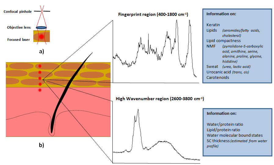
Figure 1: Measurement principle of confocal Raman microspectroscopy. a) Monochromatic laser light is focused in the stratum corneum (SC) or deeper epidermis. The photons originating from the very focus of the laser beam are detected, where a confocal pinhole assures rejection of out-of-focus light and high resolution in depth. b) The photons, which undergo frequency shifts due to the release of energy to molecules during the interaction, are used to obtain the Raman spectra. Each spectrum is derived from a point-measurement. Raman images can be obtained by scanning a laser beam over an area or displacing an object under a stationary laser beam. The position and intensity of each peak are representative of the different molecules and their amounts, respectively. Raman spectra can be obtained in a low (fingerprint) or high energetic region: each region contains different information about the molecular composition. Reprinted with permission from D. Falcone et al., Skin Pharmacol. Physiol., 28, 307-317 (2015). Copyright (2015), S. Karger AG, Basel.