
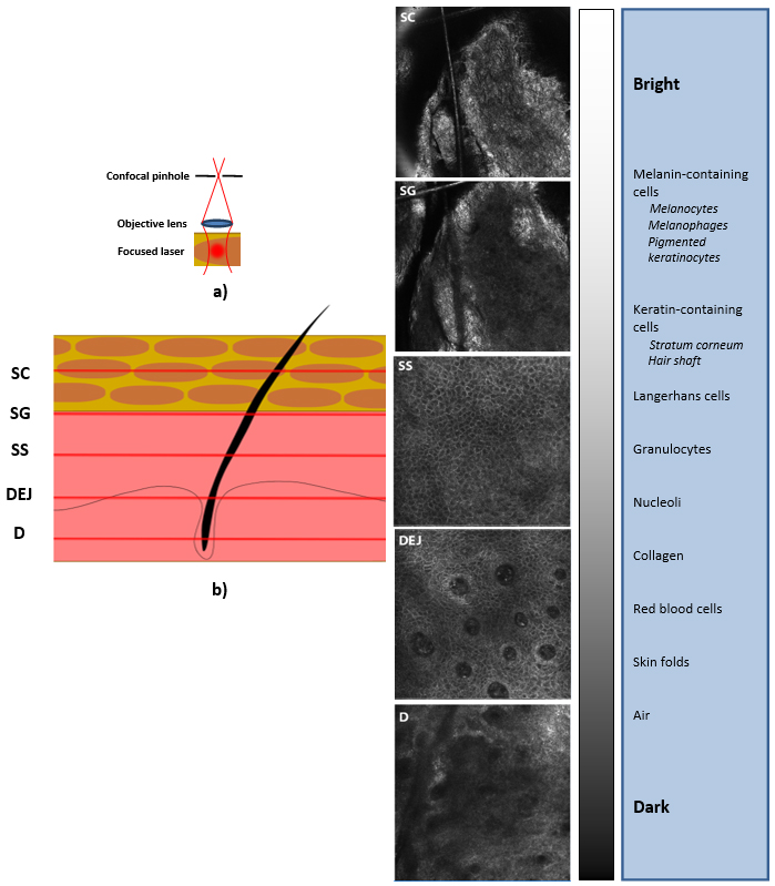
Figure 3: Measurement principle of reflectance confocal microscopy. a) Monochromatic laser light is focused in in the stratum corneum (SC) or deeper epidermis and dermis. The photons originating from the varying focus points of the laser beam are detected, where a confocal pinhole assures rejection of out-of-focus light and high resolution in depth. b) The photons which are reflected from the skin are used to obtain the reflectance confocal microscopy images. Each image is typically obtained by combining several point-measurements of the laser light through the use of a scanning optics. The intensity of the gray-level images is representative of the refractive index of different structures in the skin: structures with a higher refractive index appear bright. The SC appears gray and no nuclei are observed. The stratum granulosum (SG) and stratum spinosum (SS) can be recognized by the appearance of dark round nuclei with a surrounding bright cytoplasm in a regular honeycomb pattern. At the dermal-epidermal junction (DEJ), the dermal papillae are visible as dark round spots. The dermis (D) appears dark gray, with a loosened, fiber-like network. Modified from Malou Peppelman, In Vivo Reflectance Confocal Microscopy: Innovations in Skin Imaging, Ph.D. thesis, Radboud University of Nijmegen, the Netherlands (2015).