
Denise Falcone,1Malou Peppelman,1Piet van Erp,1Anna Ezerskaya,3Babu Varghese,2 and Natallia E. Uzunbajakava2
1Radboud University Medical Center, Department of Dermatology, René Descartesdreef 1, 6500 HB, The Netherlands
2Philips Research, High Tech Campus 34, 5656 AE Eindhoven, The Netherlands
3Optics Research Group, Delft University of Technology, Delft 2628 CH, The Netherlands
Introduction
In the era of 'Quantified Self',1 logging data about our behavior, health, and physical and mental performance has become part of our daily routine.
From an anatomic and physiological perspective, human skin represents 'a complete biologic universe'. Not only does it house the skin appendages, such as sweat glands and pilosebaceous units, but also blood vessels, muscle tissue, nerves, components of immuno-competence and endocrine function, and more.2 Furthermore, research over the past ten years has demonstrated the skin's remarkable stress sensing capacity.3 The function of human skin thus goes far beyond a 'guardian' of water-holding capacity and mechanical integrity. The skin participates in the homeostasis of the entire body.
The most 'traditional' function of the skin, the barrier function, is carefully regulated by proliferation of epidermal keratinocytes and by their subsequent progressive well-orchestrated differentiation. In this process of formation of the ultimate 'barrier', keratinocytes gradually undergo a transformation into highly cohesive corneocytes, accompanied by the synthesis of different types of keratins, lipids, and natural moisturizing factor (NMF).4 The immune function of the skin is defined by an interplay of immune mediators (e.g., interleukins and chemokines released by keratinocytes and fibroblasts), which, upon perturbation, prompts the onset of an inflammatory response, with recruitment of systemic immune cells (like dendritic cells, neutrophils, and T-cells).5
Several methods for evaluation of skin integrity and physiology in relation to its structure and function are well accepted in the dermatologist's office and in the mainstream of the cosmetic industry. They include histological and immunohistochemical analyses of punch biopsies—the "gold standard" for diagnosis, monitoring of skin disease, and evaluation of skin integrity and physiology. On the other hand, easy-to-use and high throughput in vivo non-invasive methods such as transepidermal water loss (TEWL), capacitance, and conductance measurements are commonly employed in the cosmetic industry to evaluate the effect of treatments.6
With a continuous inflow of new knowledge on cutaneous biology, rising evidence about cutaneous manifestations of systemic diseases,7 and high demands towards personalized-aesthetics treatments,8 there is a clear need for non-invasive, quantitative skin measurements fulfilling the growing demands of 'Quantified Self'. In this letter, we specifically focus on novel optical methods: reflectance confocal microscopy and Raman and near-infrared microspectroscopy. We will compare these techniques with traditional, mainstream approaches, and emphasize benefits they could offer for personalization of skin care solutions in terms of quantification of the skin structure, its barrier, and cutaneous inflammatory processes.
Non-invasive, In Vivo Analysis of the Skin Barrier: Established Versus Top-notch Approaches
Over the last three decades, stratum corneum barrier function and hydration have been extensively evaluated by means of TEWL and electrical methods such as capacitance and conductance.9,10 These techniques are implemented in relatively low cost, hand-held probes allowing for simple, rapid, in vivo, and non-invasive measurement of a physical or electrical parameter considered representative of the status of the skin barrier. The simplicity of these traditional, easy-to-use techniques comes, however, at a cost of their specificity. The data interpretation is not straightforward, as read-outs might be affected by external and internal factors not taken into account by these techniques. In particular, in the case of the electrical techniques, substances or treatments that interact with the keratin-water network of the stratum corneum can change the electrical properties of the skin without actually altering the water content.11 Moreover, despite the apparent ease of use, measurements have to be performed under strictly controlled conditions in order to obtain reliable results, minimizing the influence of biasing factors, such as ambient temperature and humidity as well as skin appendages.11,12
In contrast, optical methods based on light absorption and/or scattering by specific molecules, such as near-infrared and Raman microspectroscopy,13,14 are well-known for their chemical specificity and high spatial resolution and, thus, are inherently superior to traditional indirect electrical methods. In confocal Raman microspectroscopy, photons of the incident laser beam interact with distinct vibrational and rotational levels of endogenous molecules. During this process—called inelastic light scattering—a portion of the photon energy is then transferred to a molecule. Thus, the scattered photons undergo a shift in a wavelength, strictly defined by the molecular structure. Detection of this tiny shift in color, as well as the intensity of the scattered laser beam, allows probing the amount and type of molecules present in the skin, while confocal implementation of the technique assures high, sub-cellular, spatial resolution of a few micrometers.14 This top-notch methodology has been widely recognized and accepted by researchers working on the frontier of skin science as well as single cell research.15,16 The endogenous molecular components directly measurable with confocal Raman microspectroscopy are: water (including the possibility to differentiate the different bound states),14,17 lipids (including the possibility to differentiate between ceramides/fatty acids and cholesterol),15,18 and NMF.14 In addition, the thickness of the stratum corneum can be indirectly measured.19
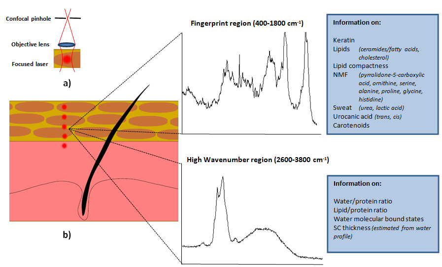
Figure 1: Measurement principle of confocal Raman microspectroscopy.
In a recent review, we investigated the relation between skin barrier assessment with indirect biophysical methods (e.g., capacitance) and direct measurements of stratum corneum composition (confocal Raman microspectroscopy) by evaluating studies in which measurements were made concomitantly by the two methods.20 Such a relationship between the two types of techniques is not always present or straightforward. For example, in a study by Crowther and co-workers, the stratum corneum water content measured with confocal Raman microspectroscopy after treatment with moisturizers containing glycerol, or a combination of glycerol and niacinamide, increased only for the latter, yet capacitance increased for both moisturizers. The authors attributed this outcome to the high dielectric constant of glycerol.21 It is clear that measuring stratum corneum composition directly, instead of measuring an integral, indirect biophysical parameter from the skin surface, allows a much more specific and complete understanding of the skin barrier. Furthermore, besides characterizing the stratum corneum molecular composition, confocal Raman microspectroscopy has also proven useful in other dermatological applications. For example, confocal Raman microspectroscopy can track the in vivo penetration of actives and drugs within the cornified envelope, provided the substances under investigation have a Raman signal, and the amount applied is sufficient to be detected by currently available devices.22 This technique can also discriminate between neoplastic and normal skin tissue, as well as between melanoma and benign melanocytic lesions.23,24
Bringing Raman Spectroscopy in the Mainstream of Clinical and Dermatological Practice
Despite the proven potential, confocal Raman microspectroscopy still has a way to go before entering into the mainstream of clinical and dermatological practice. Current state-of-the-art in vivo confocal Raman microspectroscopy applications demand a high-performance light source and detection optics as well as specific software and 'know-how' to process and extract the information of interest,15,25,26 despite past attempts to simplify the technique and lower its costs.27 A possible approach to reduce size, cost, and complexity could exploit the fact that relevant diagnostic information is often contained in a limited number of spectral regions. This would allow for the development of dedicated Raman devices for specific applications. Instead of using conventional optical components, integrated optics technology could be used in which the components are miniaturized and mass-produced.25 However, in order to develop such applications and ultimately achieve broader use of confocal Raman microspectroscopy, a close collaboration between spectroscopists, dermatologists, and skin biologists is required.20
Near-infrared Microspectroscopy: Quantification of Water and Lipids at Lower Complexity and Costs
Near-infrared microspectroscopy is an alternative approach to confocal Raman microspectroscopy for quantification of the molecular composition of the skin, since this technique offers lower complexity and cost due to less demanding laser and detection optics. Although having far less molecular specificity compared to Raman scattering, near-infrared microspectroscopy can provide quantitative and molecular-specific information on water and lipids in the skin—two components that play an important role in skin condition (e.g., oily skin versus dry skin) as well as skin barrier and its disorders.4 We have recently demonstrated a novel non-invasive near-infrared microspectroscopic technique for simultaneous measurement of oiliness and hydration levels of the skin.28 The technique utilizes differential detection with three wavelengths (1720, 1750, and 1770 nm) corresponding to the lipid vibrational bands positioned between the prominent water absorption bands. Besides lower cost, another advantage of the proposed method over confocal Raman microspectroscopy is that the measurement probe does not need to be in contact with the skin, so that repeated measurements can be performed on the same location without changing the skin conditions. Quantitative and simultaneous determination of water and lipids should enable clinicians to classify the skin types as normal, dry, oily, oily-hydrated, and oily-dry skin in a more reliable manner as compared to traditional techniques, and could provide more accurate personalized skin treatment solutions.
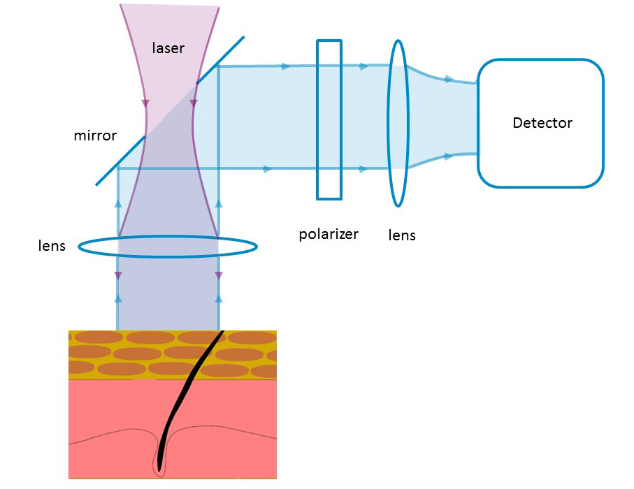
Figure 2: Measurement principle of near-infrared spectroscopy. Skin is illuminated with narrowband polarized near infrared light. Skin hydration and sebum levels are estimated from the back-reflected light using a detector in cross-polarized mode.
Reflectance Confocal Microscopy: In Vivo Imaging of Inflammatory Processes in the Skin
While confocal Raman and near-infrared microspectroscopy can directly measure the molecular composition of the skin, they neither allow direct assessment of the skin morphology (thickness of the epidermis as well as the size and shape of the epidermal-dermal junction, pilosebaceous unit, and upper plexus vasculature) nor of the immune response (e.g., the presence of inflammatory cell infiltration in inflammatory skin diseases such as psoriasis vulgaris and alopecia areata).29,30
For this purpose, another technique has been successfully used in dermatology and cosmetic science: reflectance confocal microscopy. In this non-invasive, in vivo skin imaging technique the contrast is provided by refractive index differences between the cell structures and the surrounding tissue. Melanin and keratin are the major contributors to such contrast in the images, while the reflectivity of white blood cells, chromatin, collagen, and elastin is involved to a lesser extent.31-34
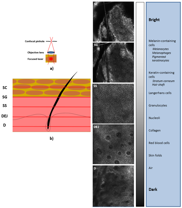
Figure 3: Measurement principle of reflectance confocal microscopy.
In clinical dermatology, successful attempts have been described in the literature for using reflectance confocal microscopy to diagnose skin cancer and inflammatory skin diseases such as psoriasis,35,36 as well as the possibility to evaluate the dynamics of neutrophil accumulation in psoriatic lesions.30 In addition, reflectance confocal microscopy is a useful tool for monitoring dynamic changes, such as tissue growth, wound healing, lesion progression, and therapy follow-up.37-42 In these clinical situations, reflectance confocal microscopy offers several advantages over conventional histology. Imaging is painless and non-invasive, causing no tissue damage. Interpretive ambiguities are reduced, since the skin is not altered by tissue processing (fixation, sectioning, and mounting) or staining, which could cause disruption of the native structure of the skin. Moreover, an inflammatory response is prevented, which may possibly interfere with the diagnosis and study observations. Further, real-time data collection is faster than routine histology and the same location of the skin can be repeatedly imaged over time.
All these advantages are also valuable in more fundamental studies on the skin barrier function and skin irritation. We have shown that tape-stripping, in combination with reflectance confocal microscopy, can be used as an in vivo model to study mechanical skin damage.43 Remarkably, a strong correlation was found between the thicknesses of the stratum corneum and living epidermis, as measured by conventional histology and reflectance confocal microscopy. We concluded that epidermal thickness assessed by reflectance confocal microscopy can be used as a measure for keratinocyte proliferation in vivo. In another in vivo model, in which the topical application of leukotriene B4 (LTB4) was used to induce a local skin inflammatory response, we were able to follow the dynamics of polymorphonuclear leukocytes migration from the dermis to the stratum corneum.44 In these models, reflectance confocal microscopy could also visualize parakeratosis, epidermal atypia, and spongiosis. These findings show that reflectance confocal microscopy imaging might be used as a non-invasive tool to obtain in vivo morphological data on material-skin barrier interactions and on the accompanying inflammatory processes.
Summary and Perspective
It is difficult to overestimate the benefits that confocal Raman microspectroscopy, near-infrared microspectroscopy, and reflectance confocal microscopy can offer for personalization of skin care solutions. Reflectance confocal microscopy is already on its way towards implementation in clinical dermatology.45 A randomized controlled trial is currently ongoing at the dermatology department of Radboud University Medical Center in Nijmegen, the Netherlands, in which reflectance confocal microscopy is being investigated as a tool for in the diagnosis of basal cell carcinoma.46 Confocal Raman microspectroscopy and near-infrared microspectroscopy could offer even more specific diagnostic information. It is the hope that this would lead to evidence-based choice of treatment for skin diseases in which skin barrier disruption plays a role (e.g., psoriasis and atopic dermatitis) as well as evaluate the skin barrier when impairment or effects of treatments are subtle (e.g., sensitive skin or following use of mild personal care devices and cosmetic applications). Ultimately, an in vivo and non-invasive approach in dermatology and skin research would help to pave the path towards personalized treatments based on differences in structure, molecular composition and cutaneous immunological status of individuals.
References
1. M. Swan, The quantified self: Fundamental disruption in big data science and biological discovery, Big Data, 1, 85-99 (2013).
2. D.J. Tobin, Introduction to skin aging, J. Tissue Viability, Mar 14 (2016); doi: 10.1016/j.jtv.2016.03.002.
3. P.C. Arck, A. Slominski, T.C. Theoharides, E.M. Peters, and R. Paus, Neuroimmunology of stress: skin takes center stage, J. Invest. Dermatol., 126, 1697-1704 (2006).
4. K.C. Madison, Barrier function of the skin: "La raison d'etre" of the epidermis, J. Invest. Dermatol., 121, 231-241 (2003).
5. G.N. Stamatas, A.P. Morello, and D.A. Mays, Early inflammatory processes in the skin, Curr. Mol. Med., 13, 1250-1269 (2013).
6. R. Darlenski, S. Sassning, N. Tsankov, and J.W. Fluhr, Non-invasive in vivo methods for investigation of the skin barrier physical properties, Eur. J. Pharm. Biopharm., 72, 295-303 (2009).
7. J.P. Callen, J.L. Jorizzo, J.J. Zone, W. Piette, M.A. Rosenbach, and R.A. Vleugels, Dermatological Signs of Systemic Disease, 5th ed., Elsevier Health Sciences (2016).
8. A.E. Rizzo and H.I. Maibach, Personalizing dermatology: the future of genomic expression profiling to individualize dermatologic therapy, J. Dermatolog. Treat., 23, 161-167 (2012).
9. G.E. Nilsson, Measurement of water exchange through skin, Med. Biol. Eng. Comput., 15, 209-218 (1977).
10. H. Tagami, M. Ohi, K. Iwatsuki, Y. Kanamaru, M. Yamada, and B. Ichijo, Evaluation of the skin surface hydration in vivo by electrical measurement, J. Invest. Dermatol., 75, 500-507 (1980).
11. E. Berardesca, European Group for Efficacy Measurements on Cosmetics and Other Topical Products, EEMCO guidance for the assessment of stratum corneum hydration: electrical methods, Skin Res. Technol., 3, 126-132 (1997).
12. V. Rogiers and E. Group, EEMCO guidance for the assessment of transepidermal water loss in cosmetic sciences, Skin Pharmacol. Appl. Skin Physiol., 14, 117-128 (2001).
13. E.M. Attas, M.G. Sowa, T.B. Posthumus, B.J. Schattka, H.H. Mantsch, and S.L. Zhang, Near-IR spectroscopic imaging for skin hydration: the long and the short of it, Biopolymers, 67, 96-106 (2002).
14. P.J. Caspers, G.W. Lucassen, E.A. Carter, H.A. Bruining, and G.J. Puppels, In vivo confocal Raman microspectroscopy of the skin: noninvasive determination of molecular concentration profiles, J. Invest. Dermatol., 116, 434-442 (2001).
15. P.D. Pudney, E.Y. Bonnist, P.J. Caspers, J.P. Gorce, C. Marriot, G.J. Puppels, S. Singleton, and M.J. van der Wolf, A new in vivo Raman probe for enhanced applicability to the body, Appl. Spectrosc., 66, 882-891 (2012).
16. N. Uzunbajakava, A. Lenferink, Y. Kraan, E. Volokhina, G. Vrensen, J. Greve, and C. Otto, Nonresonant confocal Raman imaging of DNA and protein distribution in apoptotic cells, Biophys. J., 84, 3968-3981 (2003).
17. R. Vyumvuhore, A. Tfayli, H. Duplan, A. Delalleau, M. Manfait, and A. Baillet-Guffroy, Effects of atmospheric relative humidity on stratum corneum structure at the molecular level: ex vivo Raman spectroscopy analysis, Analyst, 138, 4103-4111 (2013).
18. M. Janssens, J. van Smeden, G.J. Puppels, A.P. Lavrijsen, P.J. Caspers, and J.A. Bouwstra, Lipid to protein ratio plays an important role in the skin barrier function in patients with atopic eczema, Br. J. Dermatol., 170, 1248-1255 (2014).
19. A. Bohling, S. Bielfeldt, A. Himmelmann, M. Keskin and K.P. Wilhelm, Comparison of the stratum corneum thickness measured in vivo with confocal Raman spectroscopy and confocal reflectance microscopy, Skin Res. Technol., 20, 50-57 (2014).
20. D. Falcone, N.E. Uzunbajakava, B. Varghese, G.R. de Aquino Santos, R.J. Richters, P.C. van de Kerkhof, and P.E. van Erp, Microspectroscopic confocal Raman and macroscopic biophysical measurements in the in vivo assessment of the skin barrier: Perspective for dermatology and cosmetic sciences, Skin Pharmacol. Physiol., 28, 307-317 (2015).
21. J.M. Crowther, A. Sieg, P. Blenkiron, C. Marcott, P.J. Matts, J.R. Kaczvinsky, and A.V. Rawlings, Measuring the effects of topical moisturizers on changes in stratum corneum thickness, water gradients and hydration in vivo, Br. J. Dermatol., 159, 567-577 (2008).
22. J. Lademann, M.C. Meinke, S. Schanzer, H. Richter, M.E. Darvin, S.F. Haag, J.W. Fluhr, H.J. Weigmann, W. Sterry, and A. Patzelt, In vivo methods for the analysis of the penetration of topically applied substances in and through the skin barrier, Int. J. Cosmet. Sci., 34, 551-559 (2012).
23. A. Nijssen, S. Koljenovic, T.C. Bakker Schut, P.J. Caspers, and G.J. Puppels, Towards oncological application of Raman spectroscopy, J. Biophotonics, 2, 29-36 (2009).
24. I.P. Santos, P.J. Caspers, T.C. Bakker Schut, R. van Doorn, V. Noordhoek Hegt, S. Koljenovic, and G.J. Puppels, Raman spectroscopic characterization of melanoma and benign melanocytic lesions suspected of melanoma using high-wavenumber Raman spectroscopy, Anal. Chem., 88, 7683-7688 (2016).
25. A.C. Baclig, N. Ismail, R.M. de Ridder, M. Pollnau, P.J. Caspers, and G.J. Puppels, Low-resolution Raman spectroscopy over a wide spectral range with a single-diffraction order arrayed-waveguide grating, J. Raman Spectr., 43, 1306-1311 (2012).
26. A. van der Pol and P.J. Caspers, "Confocal Raman Spectroscopy for In Vivo Skin Hydration Measurement" in Handbook of Cosmetic Science and Technology, 3rd ed., Eds. A.O. Barel, M. Paye, and H.I. Maibach, Informa Healthcare: New York (2009).
27. N. Uzunbajakava, P. de Peinder, G.W. t'Hooft, and A.T. van Gogh, Low-cost spectroscopy with a variable multivariate optical element, Anal. Chem., 78, 7302-7308 (2006).
28. A. Ezerskaia, S.F. Pereira, H.P. Urbach, R. Verhagen, and B. Varghese, Quantitative and simultaneous non-invasive measurement of skin hydration and sebum levels, Biomed. Opt. Express, 7, 2311-2320 (2016).
29. M. Ardigo, M. Agozzino, C. Franceschini, C. Donadio, L.S. Abraham, L. Barbieri, I. Sperduti, E. Berardesca, and S. González, Reflectance confocal microscopy for scarring and non-scarring alopecia real-time assessment, Arch. Dermatol. Res., 308, 309-318 (2016).
30. E.A. Wolberink, M. Peppelman, P.C. van de Kerkhof, P.E. van Erp, and M.J. Gerritsen, Establishing the dynamics of neutrophil accumulation in vivo by reflectance confocal microscopy, Exp. Dermatol., 23, 184-188 (2014).
31. P. Calzavara-Pinton, C. Longo, M. Venturini, R. Sala, and G. Pellacani, Reflectance confocal microscopy for in vivo skin imaging, Photochem. Photobiol., 84, 1421-1430 (2008).
32. D.S. Gareau, Y.G. Patel, and M Rajadhyaksha, "Basic principles of reflectance confocal microscopy" in Reflectance Confocal Microscopy of Cutaneous Tumors, Eds. S. González , M. Gill, and A.C. Halpern, Informa Healthcare: London (2008).
33. M. Rajadhyaksha, S. González, J.M. Zavislan, R.R. Anderson, and R.H. Webb, In vivo confocal scanning laser microscopy of human skin. II: Advances in instrumentation and comparison with histology, J. Invest. Dermatol., 113, 293-303 (1999).
34. M. Rajadhyaksha, M. Grossman, D. Esterowitz, R.H. Webb, and R.R. Anderson, In vivo confocal scanning laser microscopy of human skin: melanin provides strong contrast, J. Invest. Dermatol., 104, 946-952 (1995).
35. L. Hoogedoorn, M. Peppelman, P.C. van de Kerkhof, P.E. van Erp, and M.J. Gerritsen, The value of in vivo reflectance confocal microscopy in the diagnosis and monitoring of inflammatory and infectious skin diseases: A systematic review, Br. J. Dermatol., 172, 1222-1248 (2015).
36. M. Peppelman, E.A. Wolberink, W.A. Blokx, P.C. van de Kerkhof, P.E. van Erp, and M.J. Gerritsen, In vivo diagnosis of basal cell carcinoma subtype by reflectance confocal microscopy, Dermatol., 227, 255-262 (2013).
37. S. González and Z. Tannous, Real-time, in vivo confocal reflectance microscopy of basal cell carcinoma, J. Am. Acad. Dermatol., 47, 869-874 (2002).
38. E. Richtig, V. Ahlgrimm-Siess, S. Koller, A. Gerger, M. Horn, J. Smolle, and R. Hofmann-Wellenhof, Follow-up of actinic keratoses after shave biopsy by in-vivo reflectance confocal microscopy—a pilot study, J. Eur. Acad. Dermatol. Venereol., 24, 293-298 (2010).
39. M. Ulrich, D. Krueger-Corcoran, J. Roewert-Huber, W. Sterry, E. Stockfleth, and S. Astner, Reflectance confocal microscopy for noninvasive monitoring of therapy and detection of subclinical actinic keratoses, Dermatol., 220, 15-24 (2010).
40. M. Venturini, R. Sala, S. González, and P.G. Calzavara-Pinton, Reflectance confocal microscopy allows in vivo real-time noninvasive assessment of the outcome of methyl aminolaevulinate photodynamic therapy of basal cell carcinoma, Br. J. Dermatol., 168, 99-105 (2013).
41. E.A. Wolberink, P.E. van Erp, R.T. de Boer-van Huizen, P.C. van de Kerkhof, and M.J. Gerritsen, Reflectance confocal microscopy: an effective tool for monitoring ultraviolet B phototherapy in psoriasis, Br. J. Dermatol., 167, 396-403 (2012).
42. E.A. Wolberink, P.E. van Erp, M.M. Teussink, P.C. van de Kerkhof, and M.J. Gerritsen, Cellular features of psoriatic skin: imaging and quantification using in vivo reflectance confocal microscopy, Cytometry B Clin. Cytom., 80, 141-149 (2011).
43. M. Peppelman, W.A. van den Eijnde, E.J. Jaspers, M.J. Gerritsen, and P.E. van Erp, Combining tape stripping and non-invasive reflectance confocal microscopy: an in vivo model to study skin damage, Skin Res. Technol., 21, 474-484 (2015).
44. M. Peppelman, E.A. Wolberink, M.J. Gerritsen, P.C. van de Kerkhof, and P.E. van Erp, Application of leukotriene B4 and reflectance confocal microscopy as a noninvasive in vivo model to study the dynamics of skin inflammation, Skin Res. Technol., 21, 232-240 (2015).
45. L. Hoogedoorn, M.J. Gerritsen, E.A. Wolberink, M. Peppelman, P.C. van de Kerkhof, and P.E. van Erp, A four-phase strategy for the implementation of reflectance confocal microscopy in dermatology, J. Eur. Acad. Dermatol. Venereol., 30, 1308-1314 (2016).
46. M. Peppelman, K.P. Nguyen, H.A. Alkemade, B. Maessen-Visch, J.C. Hendriks, P.E. van Erp, E.M. Adang, and M.J. Gerritsen, Diagnosis of basal cell carcinoma by reflectance confocal microscopy: Study design and protocol of a randomized controlled multicenter trial, JMIR Res. Protoc., 5, e114 (2016).